Popular Science - Is It a Pool of Moonlight or a Hoar-Frost on the Ground?
2024-09-12 09:00:17
Popular Science - Is It a Pool of Moonlight or a Hoar-Frost on the Ground?
On a tranquil night in the fifteenth year of the Kaiyuan era during the Tang Dynasty, Li Bai, slightly intoxicated, gazed upon the bright moon in the sky. Facing the moonlight that poured down like water and cast a clear radiance, the poet's longing for his hometown arose spontaneously, and he could not help but say, "Before my bed a pool of light—Can it be hoar-frost on the ground?" This left future generations with this everlasting famous line. Why did the poet associate the "moonlight" with "frost on the ground" in his slight drunkenness? This involves the mechanism of visual formation in the brain. Let us accompany the poet's talent to the threshold of neuroscience and explore the mysteries of vision.
- The Amazing Brain
The brain is one of the most essential organs in the human body and the most complex. Human higher activities such as learning, memory, and language communication are all inseparable from the brain. It can be said that the existence of this organ has created the brilliant civilization of humankind to date. The human brain is composed of connections between approximately 10^11 to 10^14 neurons. Due to the functional similarities between neurons in the human brain and diodes, people often draw analogies between the human brain and computers. Theoretically speaking, all the neural synapses in the human brain allow the brain to perform 10^17 calculations per second, with a computing power of about 10,000 TPOS, far higher than that of an ordinary personal computer. At the same time, the human brain also has the characteristic of low energy consumption. Taking AlphaGo, which played against Lee Sedol, as an example, AlphaGo needed more than 1,000 CPUs and 176 GPUs to work simultaneously, with a power of more than 100kW. However, the power of the human brain is only 20W, far less than the former.
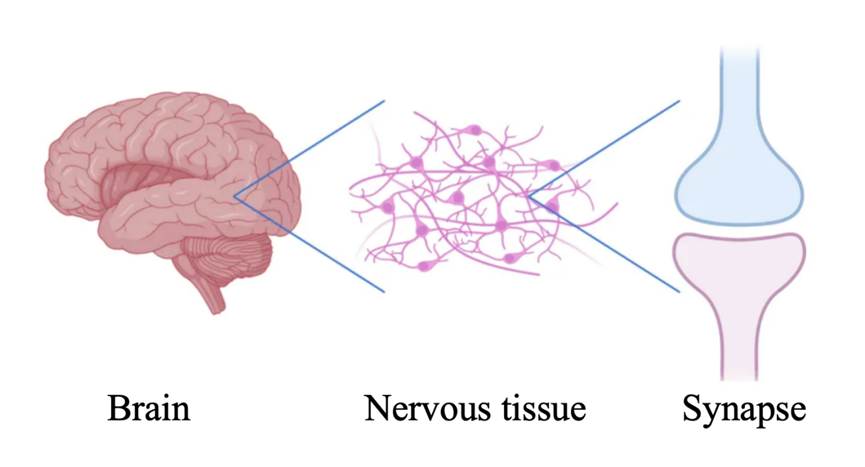
Figure 1 Schematic diagram of the brain, neural tissue, and synapses
The mechanism of vision formation we are going to talk about today is closely related to the brain. When the external scene is reflected into our eyes through light, it is first preliminarily decomposed and processed in the retina. Then, the signal is transmitted to the primary visual cortex for intermediate processing. And finally, it enters the higher center for the final integration, thus forming our vision. Now, let us embark on a journey to reveal the mysteries of brain vision together!
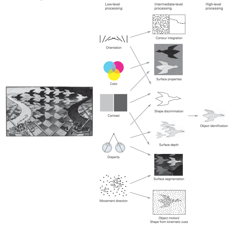
Figure 2 Hierarchical processing model of visual information
The brain processes visual scenes at low, intermediate, and high levels. Primary processing involves simple discrimination of visual attributes such as local contrast, orientation, color, and motion; intermediate processing consists of analyzing the layout of the scene and surface features, analyzing the visual image from surfaces and overall contours, and distinguishing between foreground and background; the high-level centers carry out the final integrative processing to form vision.
(The image is referenced from Principles of Neural Science, Sixth Edition, page 500)
- The Poetry Between Light and Shadow
Please close your eyes and imagine the scene Li Bai saw on an autumn night a thousand years ago: the poet looked down and saw the mottled moon shadow on the ground, and the cold moonlight appeared exceptionally bright against the backdrop of the night, which was so confusing that it was mistaken for a layer of thick frost on the ground. The boundary between light and shadow created an illusion for the poet and inspired endless poetry. If our visual system could not sensitively distinguish between light and dark, perhaps the poet could not have left behind such a beautiful verse. Why can our visual system perceive and analyze the contrast of light and shadow? Before answering this question, let us play a few interesting games.
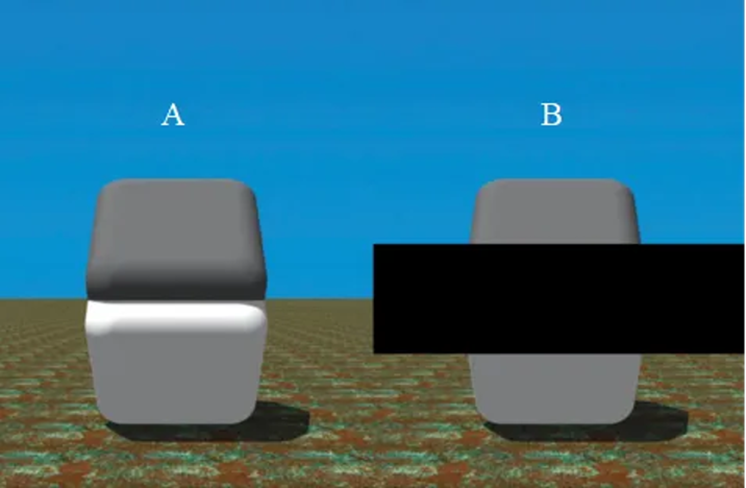
Figure 3 This is a classic example of the Cornsweet illusion.
When we observe the A shape, we tend to mistakenly believe that the upper part and the lower part have different colors. However, when we cover the area where the upper and lower parts meet (that is, the B shape), we will find that the colors of these two parts are precisely the same.
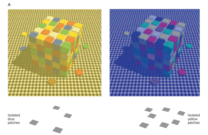
Figure 4 In the squares on the left and right, we see "yellow" small color blocks, and it is easy to think that the colors of the two are the same, but this is actually a visual illusion. When we separate the "blue" small color block in the square on the left and the "yellow" small color block in the square on the right and place them on a white background for observation, we will find that these small color blocks that looked different in color are actually the same.
(The picture is cited from Principles of Neural Science, Sixth Edition, page 557)
Figure 5 The Hermann grid comprises crisscrossing white strips and black squares. You can feel the looming gray patches at the intersection of the white strips, but in fact, the color at the intersection is white, and there are no gray patches.
(The picture is cited from Principles of Neurobiology, Second Edition, page 140)
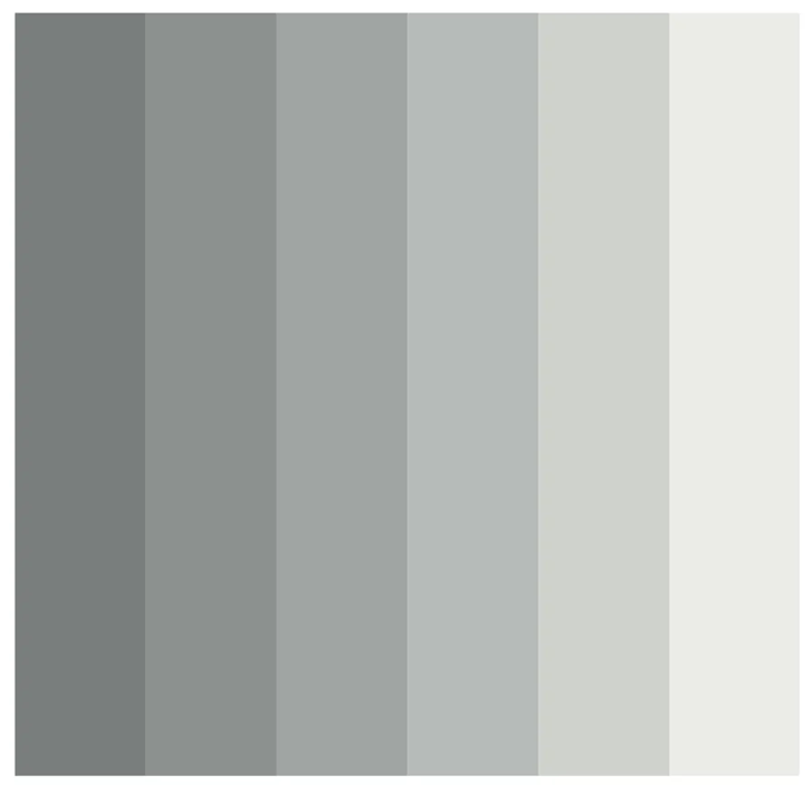
Figure 6 Mach bands are composed of a row of vertically arranged strips with different gray levels, with the gray level of the strips becoming lighter from left to right. When we observe the boundary of the strips, the color on the left appears darker, and the color on the right appears lighter. This makes it seem that the gray level of the same strip decreases from left to right, but in fact, the color in the same strip is uniform and the same.
(The picture is cited from Principles of Neurobiology, Second Edition, page 140)
The above four sets of visual games tell us that our visual system often "deceives" us, and what we see is not necessarily the truth. Many factors cause these visual illusions, one of which is that the neurons in our retina not only transmit signals of light stimulus intensity changes over time but also analyze the contrast of light and dark through the center-surround receptive field.
Scientists have found that for a ganglion cell, only light stimulation applied to a specific area of the retina can excite the cell, and we call this area the receptive field of the cell. Through experimental design, scientists have demonstrated that when the size and position of the light spot on the receptive field change, the firing of retinal ganglion cells is different. Based on this property, scientists divide ganglion cells into two types: ON-center/OFF-surround and OFF-center/ON-surround cells. For the former, when a center bright but surrounded by dark light stimulation pattern is applied, the cell's excitability is the strongest, and the latter is the opposite.
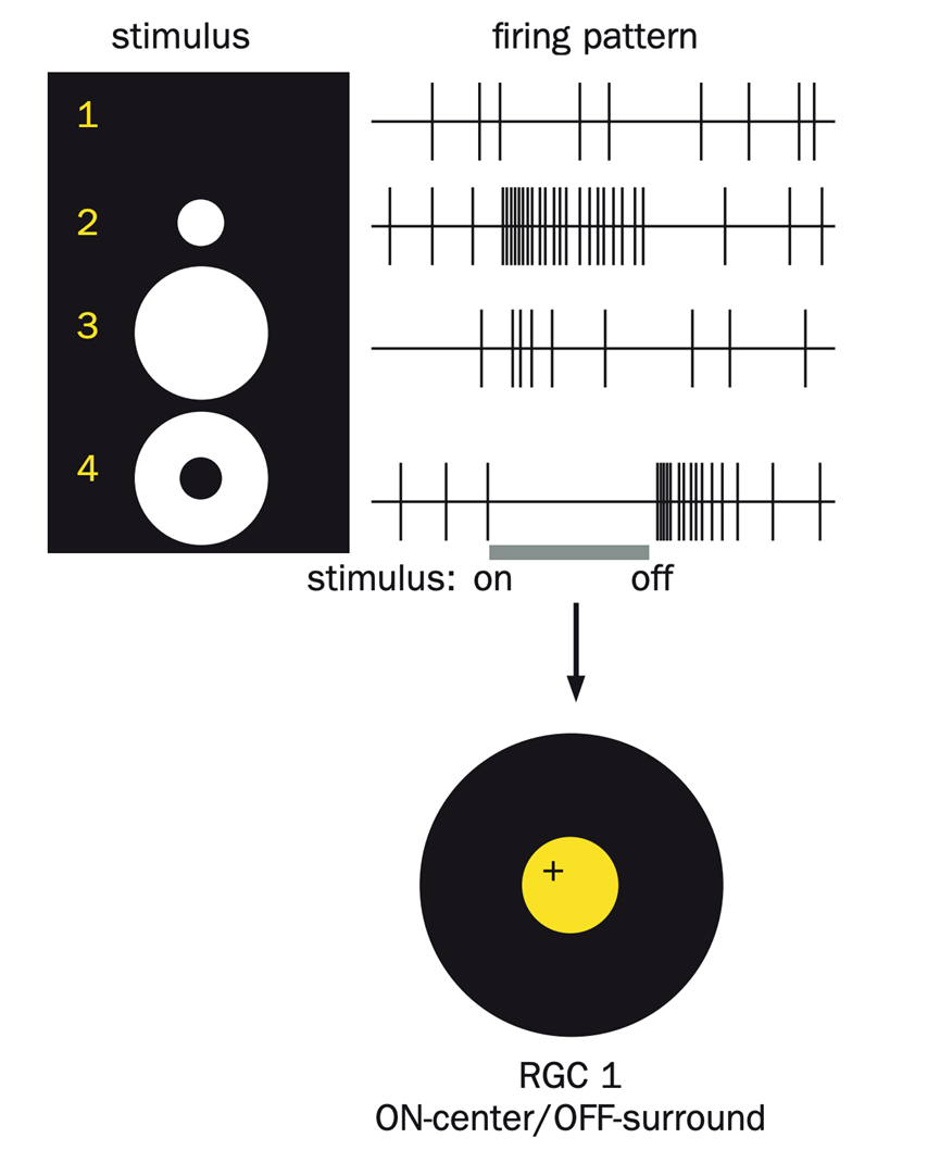
Figure 7 This is a central excitation-peripheral inhibition type neuron. Researchers applied four different stimulation patterns within the neuron's receptive field to judge its excitability based on the firing pattern. As shown in the figure, the neuron exhibits the highest level of excitability when light stimulation is applied only to the center of the receptive field (second stimulation pattern). Conversely, when light is provided in the peripheral region without illumination in the central area (fourth stimulation pattern), the neuron barely generates action potentials, indicating the lowest excitability.
(Image source: Principles of Neurobiology, Second Edition, Page 137)
This discovery explains that some cells on the retina can not only respond to light but also perform small-area retinal contrast between light and dark, thereby determining the spatial information of the light signal and enhancing our perception of the light and dark boundary. Based on this feature, we can distinguish and perceive light and dark.
- Imagination in the Haze
In addition to the perception of light and dark, the hazy atmosphere brought by night also causes the poet's uncertainty about the scene he sees, thus generating the guess of "Can it be hoar-frost on the ground?" It is this hazy sense of uncertainty that adds a subtle beauty to the poetry. If the poet were not in the dark at this time but in an environment with relatively good lighting, even if he could perceive the difference between light and dark, he might not be able to produce the excellent association between moonlight and white frost due to the improvement of the clarity of the scene. From this, we naturally think of: Why the clarity of seeing things at night decreases? To solve this problem, let us first understand the basic structure of the retina. The retina includes the pigment epithelial layer and the neural layer. Although the thickness of the retina is only 0.1-0.5 millimeters, its structure is very complex. The neural layer of the retina mainly contains photoreceptor cells, as well as bipolar cells, ganglion cells, horizontal cells, and amacrine cells, four types of neurons.
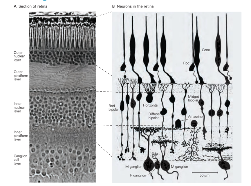
Figure 8 The retina comprises different types of neurons
(The picture is cited from Principles of Neural Science, Sixth Edition, page 523)
Among them, photoreceptor cells can be divided into two major categories: rod and cone cells, which significantly differ in cell morphology and physiological function. From Figure 9, we can see that the outer segment of rod cells is cylindrical, and the outer segment contains many overlapping disc membranes, making rod cells very sensitive to light stimulation. In contrast, the outer segment of cone cells is conical and responsible for high-acute vision and color perception functions.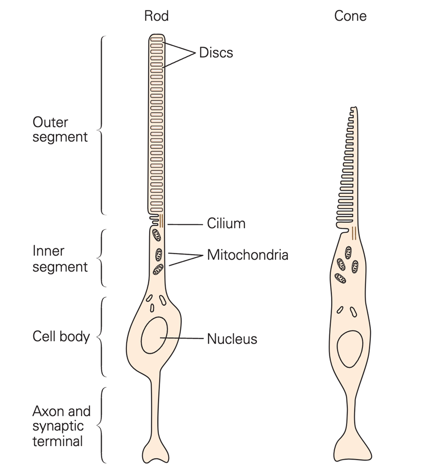
Figure 9 Rod and cone photoreceptors have similar structures
(The picture is cited from Principles of Neural Science, Sixth Edition, page 525)
Due to the high sensitivity of rod cells to light, they can perceive weak light stimulation in dim environments and cause vision. However, rod cells only contain one type of photopigment, rhodopsin, so they cannot perceive color, and their ability to distinguish the details of the object being viewed is also low; in contrast, the photosensitivity of cone cells is much weaker, but the cone cells of primates can be divided into three types according to the different photopigments they contain blue cone cells, green cone cells, and red cone cells. The different types of cone cells have very distinct bands of light to which they are most sensitive, and according to the trichromatic theory, when a specific wavelength of light acts on the retina, it will excite the three different types of cone cells in a particular proportion. This information is transmitted to the central nervous system, and the perception of the color of that light is produced.
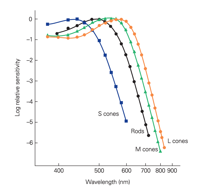
Figure 10 Correspondence between the sensitivity of different cone cells to light and wavelength.
The vertical axis is the logarithmic scale of relative sensitivity, and the horizontal axis is the wavelength of light.
S cones: Blue cone cells, M cones: Green cone cells, L cones: Red cone cells.
As depicted in the figure, different types of cone cells exhibit peak sensitivities at different wavelengths of light. Furthermore, the spectral absorption peaks of these three cone cells are close to the wavelengths of red, green, and blue light, providing evidence for the trichromatic theory.
(The picture is cited from Principles of Neural Science, Sixth Edition, page 526)
In addition, cone cells are densely arranged in the central region of the retina, which allows these cells to distinguish more minor light spots, and the central region is free of blood vessels, so light can directly stimulate these cone cells without hindrance, further strengthening high-precision vision. Up to now, we can understand why night vision will decrease. This is because the light stimulation is weak at night, and we mainly rely on rod cells to see objects clearly, but rod cells cannot produce color vision, and they are not good at dealing with high-precision vision, so our night vision is not as good as during the day.
- Conclusion
"Before my bed a pool of light—Can it be hoar-frost on the ground?" This well-known classic verse contains scientific phenomena closely related to our brain's visual system, showing that neuroscience is closely related to our daily lives. As long as we have a pair of eyes that are good at discovering, we can find scientific problems worth studying and thinking about; let us always carry a curious attitude and explore and travel in the unknown field of neuroscience.
References
1.Wang Tinghuai. Physiology. 9th Edition. Beijing: People's Medical Publishing House
2.Bear, Mark F. Neuroscience: Exploring the Brain. Lippincott Williams & Wilkins
3.Eric R. Kandel, John D. Koester. (2021). Principles of Neural Science (Sixth Edition). McGraw Hill
4.Liqun Luo. (2021). Principles of Neurobiology (Second Edition). CRC Press
Author:
Zheng Jiadong, Shanghai Medical College of Fudan University;
ZHANG Jiayi, Researcher, Institute of Brain Sciences, Fudan University,
Deputy Director of the National Key Laboratory for Brain Functions and Diseases,
Vice-Dean of the Institute of Medical-Industrial Cross-Innovation, Eye, Ear, Nose and Throat Hospital, Fudan University.
NEWS
Notice of the Shaanxi Provincial Department of Education and 16 Other Departments on Issuing the "Implementation Opinions on Strengthening Science Education in Primary and Secondary Schools in the New
The importance and significance of science and education for teenagers
The current situation of global youth science and education
Opinions on Strengthening Science Education in Primary and Secondary Schools in the New Era Issued by the Ministry of Education and 18 other Departments
Strengthen science education in primary and secondary schools in the new era! Yunnan has introduced twenty measures
International youth science practice activities and competitions
Recommendations for international science education resources

Child Friendly Cities
Introduction to CFC
News & Events
Latest Updates
Global Organizations
Organization Structure
About Us
- Introduction to UNICCC
Contact Us
Address: 8810 S GRAMERCY PL LOS ANGELES, CA 90047, U. S. A.
© United Nations Intergovernmental Cooperation Organization for the Construction of Child-Friendly Cities